Русский

Archive for A. Grevtsev

Inorg. Chem., 2018, 57, 3420-3433

A. S. Mikherdov , A. S. Novikov , M. A. Kinzhalov , V. P. Boyarskiy, G. L. Starova, A. Yu. Ivanov, V. Yu. Kukushkin

“Halides Held by Bifurcated Chalcogen-Hydrogen Bonds. Effect of µ(S,N-H)Cl Contacts on Dimerization of Cl(carbene)PdII Species ”

Inorg. Chem. , 2018, 57(6), 3420-3433
DOI:10.1021/acs.inorgchem.8b00190

 source: https://pubs.acs.org/doi/10.1021/acs.inorgchem.8b00190

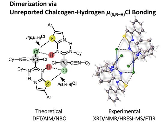
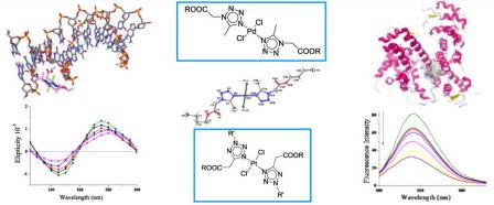
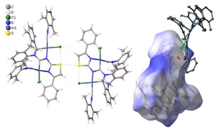
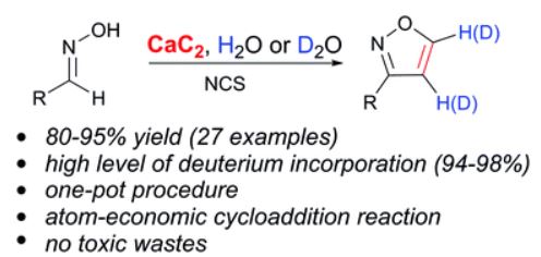
The reaction of cis-[PdCl2(CNCy)2] (1) with thiazol-2-amines (2–10) leads to the C,N-chelated diaminocarbene-like complexes [PdCl{C(N(H)4,5-R2-thiazol-2-yl)NHCy}(CNCy)] (11–14; 82–91%) in the case of 4,5-R2-thiazol-2-amines (R, R = H, H (2), Me, Me (3), −(CH2)4– (4)) and benzothiazol-2-amine (5) or gives the diaminocarbene species cis-[PdCl2{C(N(H)Cy)N(H)4-R-thiazol-2-yl}(CNCy)] (15–19; 73–93%) for the reaction with 4-aryl-substituted thiazol-2-amines (R = Ph (6), 4-MeC6H4 (7), 4-FC6H4 (8), 4-ClC6H4 (9), 3,4-F2C6H3 (10)). Inspection of the single-crystal X-ray diffraction data for 15–17 and 19 suggests that the structures of all these species exhibit previously unrecognized bifurcated chalcogen–hydrogen bonding μ(S,N–H)Cl and also PdII···PdII metallophilic interactions. These noncovalent interactions collectively connect two symmetrically located molecules of 15–17 and 19, resulting in their solid-state dimerization. The existence of the μ(S,N–H)Cl system and its strength (6–9 kcal/mol) were additionally verified/estimated by a Hirshfeld surface analysis and DFT calculations combined with a topological analysis of the electron density distribution within the formalism of Bader’s theory (AIM method) and NBO analysis. The observed noncovalent interactions are jointly responsible for the dimerization of 15–19 not only in the solid phase but also in CHCl3 solutions, as predicted theoretically by DFT calculations and confirmed experimentally by FTIR, HRESI-MS, 1H NMR, and diffusion coefficient NMR measurements. Available CCDC data were processed under the new moiety angle, and the observed μ(S,E–H)Cl systems were classified accordingly to E (E = N, O, C) type atoms.

Inorganica Chimica Acta, 2018, 473, 133-144

A. V.Protas, E.A.Popova, O. V.Mikolaichuk, Y. B.Porozov, A. R.Mehtiev, I. Ott, G. V. Alekseev, N. A.Kasyanenko, R. E.Trifonov

“Synthesis, DNA and BSA binding of Pd(II) and Pt(II) complexes featuring tetrazolylacetic acids and their esters”

Inorganica Chimica Acta, 2018, 473, 133-144
DOI:10.1016/j.ica.2017.12.040

 source:https://www.sciencedirect.com/science/article/pii/S0020169317317127?via%3Dihub

Two series of palladium(II) and platinum(II) complexes featuring esters of tetrazol-1-yl and tetrazol-5-ylacetic acids {trans-[PdCl2L2] and trans-[PtCl2L2], L  =  5-methyl-1H-tetrazol-1-ylacetic acid and its ethyl, butyl, isobutyl esters (1–5); 2-R-2H-tetrazol-5-ylacetic acid and its ethyl esters, R = tBu, CH2CH2OH (6–10)} were synthesized and their binding to calf-thymus DNA (CT DNA) and bovine serum albumin (BSA) were studied by means of experimental (CD, UV, viscometry, fluorometric and electrophoretic techniques) and theoretical methods. According to the spectrophotometric data, the interaction of the metal complexes with CT DNA is observed. The significant increase of melting point of CT DNA in the presence of the metal complexes (ΔTm = 8–13 °C) indicates strong stabilization of the DNA helix. Electrophoretic studies demonstrate the ability of the metal complexes to interact with pBR322 plasmid DNA and to change its mobility. According to the data of the fluorescence quenching technique, binding with constants (Kbin) of Pd(II) complexes with BSA are in the range 0.83–4.12 × 105 L M−1. The molecular docking studies show the minor groove binding behavior of tetrazole-containing palladium(II) and platinum(II) complexes to DNA (ΔGbinding. −5.56 − −6.12 kcal/mol) and effective binding to BSA via the favored binding site Trp213 (ΔGbinding −7.2 − −7.56 kcal/mol). The complex trans-[PtCl2(2-tert-butyl-tetrazol-5-ylacetic acid)2] exhibited noticeable antiproliferative activity in two human cancer cell lines with IC50 values of 11.40 µM in HT-29 cells and 11.02 µM in MDA-MB-231 cell line.

Crystals, 2018, 8, 112

A. S. Mikherdov, A. S. Novikov, M. A. Kinzhalov, A. A. Zolotarev, V. P. Boyarskiy

“Intra-/Intermolecular Bifurcated Chalcogen Bonding in Crystal Structure of Thiazole/Thiadiazole Derived Binuclear (Diaminocarbene)PdII Complexes”

Crystals, 2018, 8(3), 112
DOI:10.3390/cryst8030112

 source: http://www.mdpi.com/2073-4352/8/3/112

The coupling of cis-[PdCl2(CNXyl)2] (Xyl = 2,6-Me2C6H3) with 4-phenylthiazol-2-amine in molar ratio 2:3 at RT in CH2Cl2 leads to binuclear (diaminocarbene)PdII complex 3c. The complex was characterized by HRESI+-MS, 1H NMR spectroscopy, and its structure was elucidated by single-crystal XRD. Inspection of the XRD data for 3c and for three relevant earlier obtained thiazole/thiadiazole derived binuclear diaminocarbene complexes (3a EYOVIZ; 3b: EYOWAS; 3d: EYOVOF) suggests that the structures of all these species exhibit intra-/intermolecular bifurcated chalcogen bonding (BCB). The obtained data indicate the presence of intramolecular S•••Cl chalcogen bonds in all of the structures, whereas varying of substituent in the 4th and 5th positions of the thiazaheterocyclic fragment leads to changes of the intermolecular chalcogen bonding type, viz. S•••π in 3a,b, S•••S in 3c, and S•••O in 3d. At the same time, the change of heterocyclic system (from 1,3-thiazole to 1,3,4-thiadiazole) does not affect the pattern of non-covalent interactions. Presence of such intermolecular chalcogen bonding leads to the formation of one-dimensional (1D) polymeric chains (for 3a,b), dimeric associates (for 3c), or the fixation of an acetone molecule in the hollow between two diaminocarbene complexes (for 3d) in the solid state. The Hirshfeld surface analysis for the studied X-ray structures estimated the contributions of intermolecular chalcogen bonds in crystal packing of 3a–d: S•••π (3a: 2.4%; 3b: 2.4%), S•••S (3c: less 1%), S•••O (3d: less 1%). The additionally performed DFT calculations, followed by the topological analysis of the electron density distribution within the framework of Bader’s theory (AIM method), confirm the presence of intra-/intermolecular BCB S•••Cl/S•••S in dimer of 3c taken as a model system (solid state geometry). The AIM analysis demonstrates the presence of appropriate bond critical points for these interactions and defines their strength from 0.9 to 2.8 kcal/mol indicating their attractive nature

April

Total in April 2877 service applications were carried out.
All together measured:

  • 2729 1H spectra
  • 421 13C spectra
  • 130 DEPT spectra
  • 69 COSY spectra
  • 53 NOESY spectra
  • 49 31P spectra
  • 115 19F spectra

323 applications were carried out.

Microporous and Mesoporous Materials, 2018, 132-142

E.A. Krylova, M.G. Shelyapina, P. Nowak, H. Harańczyk, M. Chislov, I.A. Zvereva, A.F. Privalov, M. Becher, M. Vogel, V. Petranovskii

“Mobility of water molecules in sodium- and copper-exchanged mordenites: Thermal analysis and 1H NMR study”

Microporous and Mesoporous Materials, 2018, 265, 132-142
DOI:10.1016/j.micromeso.2018.02.010

 source:https://www.sciencedirect.com/science/article/pii/S1387181118300696?via%3Dihub

Comprehensive research of water behavior in Na- and Cu-mordenites with different Na/Cu ratio was done. Several steps of dehydration process were detected and analyzed, taking into account difference in chemical composition of the samples, reaction models and corresponding kinetic equations. Activation energies for these steps were calculated. It was shown that the majority of dehydration steps for all zeolite samples studied might be associated with chemical reaction mechanism corresponding to the second order kinetic model, except for the most high-temperature step for Cu-mordenite, for which the third-order model has the higher correlation coefficient. A detailed analysis of rehydration processes was studied by proton NMR spectroscopy. The obtained results allow one to distinguish different types of water and to associate them with a certain localization of water molecules in zeolite voids: the main channel for both Na and Cu-mordenites; a side pocket of Na-mordenite; molecules coordinated with Cu2+ cations in Cu-mordenite. The diffusion measurements carried out using static field gradient NMR technique proved that the water diffusion character below 300 K is essentially intracrystalline, whereas above 300 K it becomes intercrystalline. The activation energy of intercrystalline diffusion is about 28 kJ/mol and does not depend on the Na/Cu ratio. That allows us to suppose that in the studied zeolites the intercrystalline diffusion is governed by the morphology of the sample mainly.

Microporous and Mesoporous Materials, 2018, 220-228

Y.M. Zhukov, M.G. Shelyapina, I.A. Zvereva, A.Y. Efimov, V. Petranovskii

“Microwave assisted versus convention Cu2+ exchange in mordenite”

Microporous and Mesoporous Materials, 2018, 259, 220-228
DOI:10.1016/j.micromeso.2017.10.013

 source:https://www.sciencedirect.com/science/article/pii/S1387181117306662?via%3Dihub

Sodium mordenite was Cu2+-exchanged by conventional methods at ambient temperature (accepted as 20 °C) and with microwave radiation at 100 ± 1 °C. To increase the copper content, we repeat the exchange procedure several times. Both the degree of Cu2+ exchange and the environment of the Cu2+ ions depend on the method of exchange. Neither conventional, nor microwave methods do not destruct the topology of mordenite framework. XRD pattern of the mordenite persists, but slight elastic deformation and some dealumination of the surface layer occurs. For all the studied samples, all the copper ions are in Cu2+ state, neither Cu1+ nor Cu0 were detected. All the copper ions play the role of charge-balancing counter-cations. Their placement into the interior of zeolite causes the channel contraction due to electrostatic interactions of double charged cations with [AlO4]δ– units. The increase of copper content is accompanied by increasing of number of water molecules per unit cell. In fully hydrated samples the copper cations are effectively separated from the zeolite framework and are fully surrounded by a water ligand shell.

Conference

source:http://www.kfti.knc.ru/mdmr/2018/first-circular.php
The International conference “Modern Development of Magnetic Resonance 2018” will be held in Kazan, Russia on September 24-28, 2018.

Org. Chem. Front., 2018, 226-231

M. S. Ledovskaya, K. S. Rodygina, V. P. Ananikov

“Calcium-mediated one-pot preparation of isoxazoles with deuterium incorporation”

Org. Chem. Front., 2018, 5, 226-231
DOI:10.1039/C7QO00705A

 source:http://pubs.rsc.org/en/Content/ArticleLanding/2018/QO/C7QO00705A#!divAbstract

In this work, a novel synthetic methodology for the one-pot preparation of isoxazoles directly from the reaction of calcium carbide with aldoximes is reported. Calcium carbide acts as a safe and inexpensive acetylene source and, in addition, as a source of the Ca(OH)2 base to enable the generation of nitrile oxide. Various 3-substituted isoxazoles were synthesized from the corresponding aldoximes in good yields (up to 95%) and a series of new deuterated 4,5-dideuteroisoxazoles were prepared.

School for Young Scientists

source: http://mrschool2018.from-siberia.ru/

The V International School for Young Scientists “Magnetic Resonance and Magnetic Phenomena in Chemical and Biological Physics” will be held on 15 — 20 September. More info.

Int. J. Nanomedicine, 2018, 1471-1482

M. Shevtsov, B. Nikolaev, Y. Marchenko, L. Yakovleva, N.V. Skvorzov, A. Mazur, P. Tolstoy, V. Ryzhov, G. Multhoff

“Targeting experimental orthotopic glioblastoma with chitosan-based superparamagnetic iron oxide nanoparticles (CS-DX-SPIONs)”

Int. J. Nanomedicine, 2018, 13, 1471-1482
DOI: 10.2147/IJN.S152461


Glioblastoma is the most devastating primary brain tumor of the central nervous system in adults. Magnetic nanocarriers may help not only for a targeted delivery of chemotherapeutic agents into the tumor site but also provide contrast enhancing properties for diagnostics using magnetic resonance imaging (MRI)